Foot and ankle has been shown to influence proximal joint mechanics through alterations in lower extremity kinetic chain coupling. The foot, ankle, tibia, femur, and hip function as an integrated system, whereby changes in distal joint motion may propagate proximally and affect overall movement strategy.
The subtalar joint (STJ) plays a critical role in this coupling mechanism. STJ motion influences both foot abduction and tibial internal rotation, thereby contributing to knee joint kinematics. Disruption of this relationship has been associated with altered knee mechanics and dysfunctional loading patterns (Kerr et al., 2019). Furthermore, reduced ankle dorsiflexion during functional tasks such as squatting has been associated with compensatory increases in knee valgus and altered quadriceps recruitment strategies, patterns commonly observed in individuals with patellofemoral pain (Macrum et al., 2012).
Foot posture has also been linked to proximal strength characteristics. Individuals with pes planus demonstrate reduced hip strength compared to those with neutral arch alignment, suggesting that distal foot structure and function may influence lumbopelvic–hip complex performance (Zahran et al., 2017). These findings support the concept that distal stability contributes to proximal neuromuscular control.
The directionality of this relationship, however, remains bidirectional. Chuter and Janse de Jonge (2012) highlight that while coupling between segments is well established, the primary source of dysfunction is less clear. Hip abductor weakness may permit excessive femoral and tibial internal rotation, contributing to foot pronation. Conversely, excessive pronation may induce internal rotation proximally, resulting in adaptive lengthening and reduced functional capacity of the hip abductors.
Overpronation and Lower Extremity Alignment
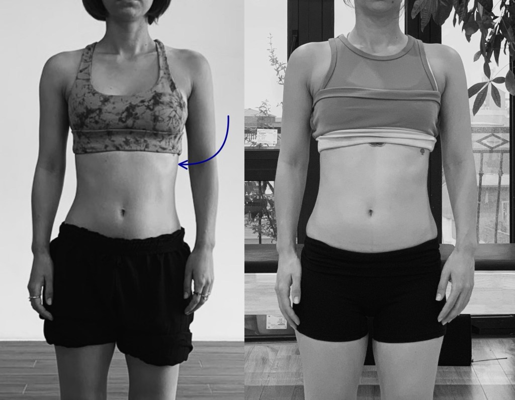
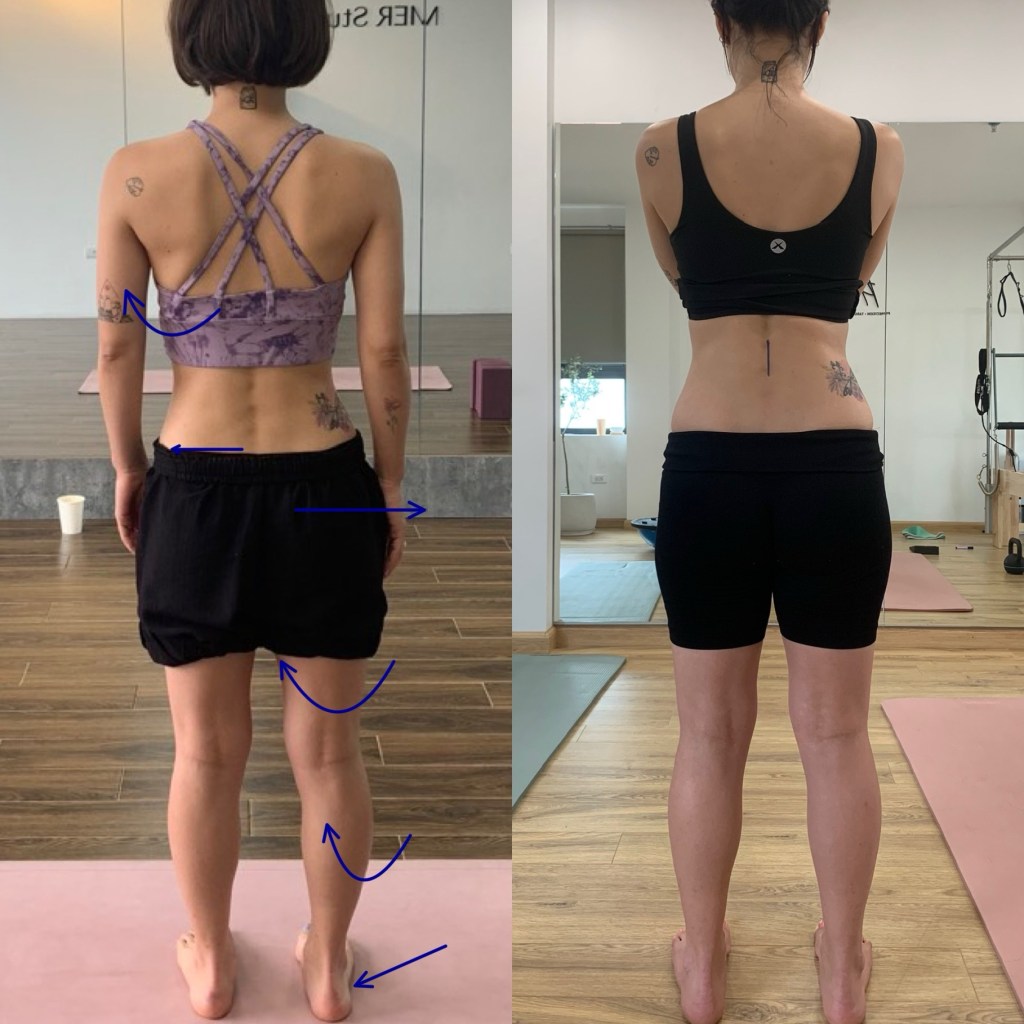
Excessive foot pronation during closed-chain activity has been associated with a characteristic kinematic chain involving tibial internal rotation, femoral adduction, and increased knee valgus (Raghava Neelapala et al., 2016). This alignment pattern reflects altered load distribution across the lower extremity and is commonly associated with dysfunctional movement mechanics.
From a neuromuscular perspective, overpronation is typically accompanied by decreased activity in key stabilizing musculature, including the tibialis anterior, tibialis posterior, and hip external rotators (gluteus maximus and medius). Concurrently, compensatory overactivity has been observed in the lateral gastrocnemius, fibularis musculature, and tensor fascia latae, suggesting a shift in motor control strategy toward less efficient synergistic patterns.
Collectively, these findings suggest that foot–ankle dysfunction may contribute to altered proximal mechanics through both kinematic coupling and neuromuscular adaptation, reinforcing the importance of integrated assessment of the lower extremity kinetic chain.
References (as cited)
- Kerr et al., 2019
- Macrum et al., 2012
- Zahran et al., 2017
- Chuter & Janse de Jonge, 2012
- Raghava Neelapala et al., 2016